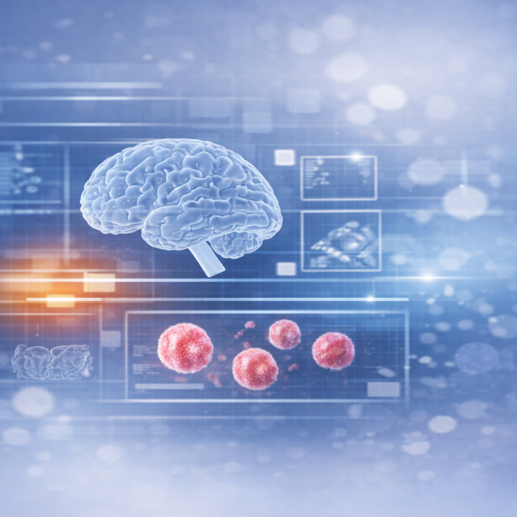
CellLogic
Our Core Architectures and Scientific Rationale
CellLogic is Cansera’s flagship technology: a two-part AI platform designed to find rare abnormal cells and deeply characterize them from blood samples. By combining unsupervised anomaly detection with single-cell phenotyping, CellLogic can flag extremely rare cellular events (like circulating tumor cells) and then classify their biological identity with high precision. This section details our core algorithms – code-named RED and BLUE – and the scientific advances that make them uniquely powerful for rare-cell analysis.
Rare Event Detection (RED): Unsupervised Anomaly Finder
The RED algorithm (Rare Event Detection) is the first stage of CellLogic, responsible for scanning a whole slide image for any cell that “looks different” from the ordinary. Unlike traditional methods that require predefined biomarkers, RED uses an unsupervised approach – it treats rare cells as anomalies in a very large pool of normal cells. An entire fluorescence whole-slide image is broken into millions of tiny image tiles (on the order of ~2.5–3 million sub-images per slide, each tile ~32×32 pixels capturing a few cells). These tiles are used to train a denoising autoencoder (DAE). Most tiles contain only normal white blood cells, so the DAE learns their patterns very well and becomes very good at reconstructing those. However, if a tile contains an unusual cell (say a tumor cell with odd morphology or marker signals), the autoencoder will reconstruct it poorly – yielding a high reconstruction error. RED thus flags these high-error tiles as potential “rare events” (i.e., candidate rare cells). This anomaly-detection strategy means we don’t have to tell RED in advance what a tumor cell looks like – the algorithm automatically learns what “normal” looks like and spots anything deviating from that norm. In practical terms, RED can sift through an entire slide unsupervised and pull out a handful of presumptive rare-cell images for further analysis – all without any human tuning. This unsupervised rare-event finder is highly generalizable: by focusing human attention only on a small, algorithm-curated subset of cells, RED massively accelerates and standardizes what was once an impossible manual task.
Single-Cell Phenotyping (BLUE): Deep Representation Learning
Once RED has isolated candidate rare cells, the BLUE pipeline takes over to identify what those cells are. BLUE is our second-stage algorithm that performs high-content single-cell segmentation and phenotypic classification. In essence, BLUE zooms into the flagged regions, extracts individual cells, and analyzes their features to determine cell type or phenotype. This involves two key steps: 1. Cell Segmentation: BLUE uses a deep learning segmentation model (based on a U-Net architecture) to precisely delineate each cell within a rare tile. This model has been trained on thousands of annotated cells to recognize cell boundaries better than conventional image processing. The segmentation ensures we isolate whole cells (distinguishing clustered cells, separating overlapping cells, etc.) for accurate analysis. 2. Feature Extraction & Classification: After segmentation, BLUE applies a representation learning approach to characterize the cell. Specifically, we use a contrastive learning framework to embed each cell’s image into a rich latent vector – effectively capturing the cell’s morphology and marker expression pattern in a numerical feature profile. Unlike traditional methods that rely on a few human-chosen features, this deep learning approach learns an unbiased, high-dimensional representation of the cell’s phenotype. Using these learned features, BLUE then classifies the cell’s identity (for example, distinguishing a circulating tumor cell from a normal white blood cell, or even subclassifying tumor cells by phenotype).
An Integrated Pipeline Fueling Discovery
The true power of CellLogic comes from integrating RED and BLUE into one seamless workflow. First, RED casts a wide net to catch anything out of the ordinary, and then BLUE investigates those catches in depth to tell us exactly what they are. This dual strategy is highly novel – it exceeds the capabilities of any single-step solution, creating a comprehensive rare-cell analysis pipeline. By eliminating reliance on predefined biomarkers, we ensure that no potential signal is ignored. By then applying state-of-the-art deep learning characterization, we ensure that no detected cell goes uninterpreted. The result is an end-to-end platform that can both discover and characterize rare cells from blood in an automated fashion. Scientifically, this approach minimizes human bias and can reveal insights that traditional methods might miss. Predefined assays tend to focus on known markers – but cancer biology is complex, and important signals can hide in unexpected cell types or marker combinations. CellLogic’s unbiased anomaly detection and rich phenotyping allow it to capture global context (the presence of any unusual cells at all) as well as fine-grained details (the phenotype of each rare cell). This opens the door to discovering novel cell subtypes or rare biomarkers that could be crucial for understanding disease progression or treatment response.